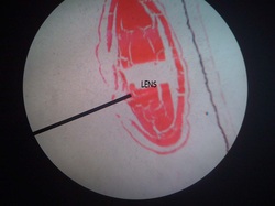
The lens consists of a lens capsule, the subcapsular epithelium and lens fibres. It does not contain blood vessels or nerves.
The lens capsule is generated by the cells of the subcapsular epithelium and corresponds to a thick, elastic basal lamina. The zonule fibres insert into the lens capsule.
Cells of the subcapsular epithelium (or anterior lens cells) are mitotically active. In adult individuals they only cover the anterior "hemisphere" of the lens. As they divide, cells gradually move towards the equator of the lens where they tranform into lens fibres. The apical part of the gradually elongating cell extends between the subcapsular epithelium and adjacent lens fibres towards the anterior pole of the lens. The basal part extends towards the posterior pole. The nucleus remains close to the equatorial plane of the lens.
The mature lens fibres, i.e. very long (up to 12 mm), hexagonal cells, form the body of the lens. They are located immediately deep to the cells of the subcapsular epithelium. Lens fibres are nucleated in the soft, outer cortex of the lens. As new lens fibres are added to the periphery of the cortex, lens fibres located deeper in the cortex loose their nuclei and become part of the somewhat harder nucleus of the lens. In the intact lens, lens fibres are tightly connected to each other. Few organelles are scattered in a cytoplasm filled withcystallin proteins. These proteins are responsible for the transparency and refractive properties of the lens and account for up to 60% of the mass of lens fibres.
Reference: www.lab.anhb.uwa.edu.au
The posterior surface of the iris is covered by the retina. The inner layer of the retina, i.e. the layer facing the posterior chamber, is called the posterior epithelium of the iris. Both layers of the retina are pigmented, but pigmentation is heavier in the inner layer. In the region of the central opening of the iris, the pupil, the retina extends for a very short distance onto the anterior surface of the iris. The iridial stroma consists of a vascularized loose connective tissue rich in melanocytes in addition to macrophages and fibrocytes, which are all surrounded by a loose meshwork of fine collagen fibers. The anterior surface of the iris is not covered by an epithelium - instead of we find a condensation of fibrocytes and melanocytes, the anterior border layer of the iris.
The iris forms the aperture of the eye. Myoepithelial cells in the outer (or anterior) layer of the retina, i.e. the layer adjacent to the stroma of the iris, have radially oriented muscular extensions. These extensions form a flat sheet immediately beneath the anterior layer of the retina, the dilator pupillae muscle. Embedded in the central portion of the iridial stroma are smooth muscle cells which form the annular sphincter pupillae muscle. In humans, this muscle surrounds the pupil as a less than 1 mm wide and only 0.2 mm thick band. The two muscles regulate the size of the pupil.
Pupillary constriction, which is mediated by the sphincter pupillae muscle, is clinically refered to as miosis - dilation, mediated by the dilator pupillae muscle, as mydriasis.
The pigmentation of cells in the stroma and anterior border layer of the iris determines to color of the eyes. If cells are heavily pigmented the eyes appear brown. If pigmentation is low the eyes appear blue. Intermediate levels create shades of green and grey.
Reference : www.lab.anhb.uwa.edu.au

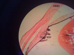
The ciliary body is an inward extension of the choroidea at the level of the lens. Ciliary processes are short extensions of the ciliary body towards the lens. A small amount of loose connective tissue similar to that of the choroid is located between smooth muscle cells which form the bulk of the ciliary body. They form three bundles, the ciliary muscle.
The inner surface of the ciliary body and its processes are lined by two layers of columnar cells which belong to the retina - the ciliary epithelium formed by the pars ciliaris of the retina. The outer cell layer is pigmented, whereas the inner cell layer, i.e. the layer that faces the posterior chamber of the eye, is nonpigmented.
The ciliary processes contain a dense network of capillaries. The cells of the inner layer of the ciliary epithelium generate the aqueous humor of the eye. , i.e. they transport the plasma filtrate generated by the capillaries in the ciliary processes into the posterior chamber of the eye. Thight junctions between the cells form the blood - aqueous humor barrier.
Fibers, which consist of fibrillin, extend from the ciliary processes towards the lens and form the suspensory ligament of the lens. These fibres are also called zonule fibres. Two of the bundles of the ciliary muscles attach to the sclera and strech the ciliary body when they contract, thereby regulating the tension of the zonule fibres. The reduced tension will result in a thickening of the lens which focusses the lens on close objects - a process called accomodation.
References : www.lab.anhb.uwa.edu.au
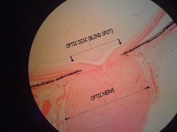
Because of the origin of the retinae and optic nerves from the developing forebrain, the optic nerves (cranial nerves II) corresponds to fibre tracts connecting parts of the CNS - in this case the ganglion cells of the retina with neurones in the lateral geniculate nucleus of the thalamus and neurones in the superior colliculus and pretectum of the midbrain.
Ganglions cell axons run towards the optic disc where they turn towards the sclera. Numerous bundles (or fascicles) of axons pass through the choroid and openings in the sclera, thelamina cribosa. The axons become myelinated in this region. Collectively, the bundles form the optic nerve. Like other parts of the CNS, the optic nerve is surrounded by the three meninges - the outer dura mater, the middle archnoid and the inner pia mater, which are separated from each other by subdural and subarachnoid spaces. At the eyeball, the dura fuses with the sclera while the arachnoid and pia mater merge with the choroid. Connective tissue septa, which arise from the pia mater, separate the fibre bundles in the optic nerve. The axons in the optic nerve are supported by astrocytes and oligodendrocytes. Microglia is also present.
Reference: www.lab.anhb.uwa.edu.au